TYPE: Research Article
Ryan Fox¹*
¹The Fox Hospital, Red Fox Rehabilitation Clinic, United Kingdom

Introduction
Red foxes (Vulpes vulpes) are known to suffer particularly in urban areas from mange, most commonly Sarcoptes scabiei (Scott et al., 2020; Soulsbury et al., 2007). However, the author has found through treating a large number of red fox patients that the underlying causes are most linked to malnutrition (mineral, nutrient, vitamin and/or amino acid deficiencies) and/or acidosis, particularly with a heavy parasite burden, which red foxes with mange commonly experience. Poor and insufficient diet through decimation of natural habitats or prey poisoned by pest control substances leading to secondary toxicity, diminished body condition through infected wounds often caused by bite wounds of cats or other foxes or open fractures and subsequent infected wounds leading to an inability to hunt effectively and a subsequently a poorly operating immune system eventually leading to a heavy parasite burden are common root causes (Fox R., unpub. data). In rural environments, this seems to be much less, likely due to various factors including but not limited to more natural and uncontaminated prey, lack of human-caused obstacles or hazardous environment and larger open space reducing the potential for territory issues (Fox R., unpub. data).
Ivermectin is commonly prescribed to treat both internal and external parasitic infections, including Sarcoptes scabiei infections. Shortly after it became available, reports of resistance to ivermectin have become widespread (Furnival-Adams et al. 2024, Laing et al. 2017, Shoop 1993). In the veterinary world, ivermectin resistance is now a concern in cases of roundworm infection in horses, cattle, and sheep, as well as canine heartworm infections (Kaplan 2004). However, reports of ivermectin resistance in human and livestock parasites have been limited and contentious to date (Crump & Ōmura 2011).
There are a number of studies pertaining to different dose schedules and ivermectin efficacy, and they appear to vary significantly in results from single dose and double dose in vitro (Mounsey et al. 2017) to 7 doses recommended by the U.S Centers for Disease Control and Prevention (CDC 2023) and U.K. National Health Service (NHS 2024). However, there does not appear to be a consensus on the optimal frequency of ivermectin administration.
The present study aimed to understand the efficacy of different dose schedules to standardise the ivermectin administration protocol for red foxes infected by Sarcoptes scabiei in the wild. The study involved, the red foxes, in vivo, in a rehab/rescue environment and in clinical best-case conditions, suggesting the accuracy to be potentially as close to ideal real-world conditions as possible.
In this study, the author refers mostly to crusted scabies since wild foxes were generally not reported by members of the public until they were advanced in their condition and already crusting. However, it was still noted that on suspicion of molting periods or possible biofilm protection, there did appear to be some ivermectin resistance based on the skin scraping results each time, resulting in a need for further doses, where the minimum amount of doses required was still above those stated in other studies.
Sarcoptes scabiei mite has three life cycle molt periods (between larvae and protonymph, protonymph and tritonymph or tritonymph and adult). Life cycle molting periods affect the efficacy of ivermectin. A study was conducted by Feng et al. (2023) demonstrated that “molting Sarcoptes mites are less susceptible to ivermectin than active mites. As a consequence, mites may survive after two doses of ivermectin given 7 days apart due not only to hatching eggs but also to the resistance of mites during their molting process. (Their) results provide insight into the optimal therapeutic regimens for scabies and highlight the need for further research on the molting process of Sarcoptes mites, where one third of mites survive the administration of ivermectin during a molting stage”.
While scabies has been observed in human patients with seemingly normal immune response, it should be noted that in red foxes, particularly adults, as demonstrated in (Supplementary Table S1), co-morbidities existed in each patient and it is the opinion of the author that there was in every case an underlying condition that appeared to have led to a reduced immune response and unnatural bodily terrain leading to the infestation of the mange mites suggesting strongly that mange is not a primary concern and instead a condition contracted and infested only once the fox was severely immunocompromised. The author notes that in his experience the exception to this appears to be only in newborns and young cubs still nursing from their already infected mother, since Sarcoptes scabiei is spread through direct contact, and the cubs then appear to become malnourished and lacking in vital nutrients via poor mother’s milk quality, leading to further decline. In the case of this report, usually either a declined immunoresponse leading to a chronic or acute parasite burden, a poorly functioning lymphatic system, previously untreated open infected wounds leading to a suspected infection of the blood, severe malnutrition and/or serious lack of essential minerals, vitamins, amino acids and nutrients due to environmental factors. The author has recognised patterns over the course of many hundreds of red fox patients treated hands-on, spanning multiple years (Fox, R., unpub. data).
Methods
A total of 111 wild red foxes (Vulpes vulpes) were rescued from 2020 to 2021, showing clinical signs and indications of decline, injury and/or mange suspicion, reported by members of the public and subsequently rescued and brought into a clinical veterinary environment for treatment. All samples were obtained as part of the standard protocol during healing therapies on rescued patients with the goal of healing and releasing them back to the wild where possible.
Skin samples from the patients were taken during the standard triage procedure within the first hour of rescue. Each time skin samples were taken by skin scrapes, a little mineral oil was placed on 3 microscope slides as well as a drop of mineral oil on the area of skin about to be sampled/scraped, then skin scrapings were carried out using a sterile size 10 scalpel blade per patient dipped in a little mineral oil (liquid paraffin) to help adhere mites and/or eggs to the blade, per patient on 3–4 areas of the body (including lesion areas, common areas on red foxes or suspected areas where it was not obvious in early stage patients. Where patients had a significant thick crust, a mineral oil-dipped dull scalpel blade or small medical spatula was used to get a sample from under the crust, with care taken to not cause the patient any pain, harm or stress. Skin was scraped on each occasion until capillary ooze was obtained to ensure removal of the epidermis where mites burrow. Skin samples from each area were then placed onto their individual microscope slides with cover slips, labelled according to the patient where necessary and the skin area sampled and examined under compound microscopy at 4x and 10x objective to identify the presence of infection (Figure 1). After samples were taken each patient was placed into isolation in either a veterinary stainless steel or polypropylene industry standard clinical veterinary kennel and remained for the duration of their treatment to avoid any cross contamination where their clinical environment could be disinfected multiple times per day as necessary to maximise the potential for the fastest healing and reduce or eliminate any possibility of environmental reinfection, with disposable incontinence pads changed frequently each day, each patient quarantined and isolated from others with no materials in kennel that could harbour any eggs, larvae or mites and in each case and each interaction, long arm length gloves and nitrile over-gloves and disposable aprons disposed of between patients and with every interaction eliminating any cross contamination. Cleaning of kennels was done using F10SCXD Veterinary Disinfectant rated for various bacteria, fungi, moulds, viruses, micro organisms and spores, and Safe4 veterinary biocide disinfectant. Another reason patients were placed into an environment capable of being disinfected multiple times per day and not into environments with hay, wood shavings, or similar and in outdoor enclosures during treatment was due to the potential for mites to fall off the host and reattach onto new hosts within 24 to 36 hours, as demonstrated by Arlian et al. (1984).
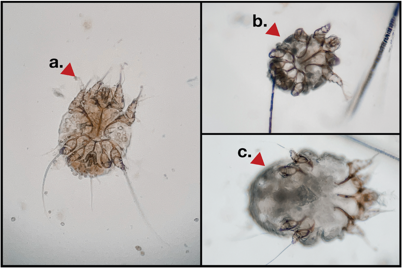
Figure 1. Sarcoptes scabiei, skin scraping samples, red foxes (Vulpes vulpes). a) ventral surface of an adult male Sarcoptes scabiei. b) ventral surface of a Sarcoptes scabiei protonymph. c) ventral surface of a pregnant adult female Sarcoptes scabiei.
Samples were taken on the initial triage day to confirm infection and again on each day of treatment, prior to treatment. The author is aware of an amended schedule which has also been implemented and recommended by the CDC following a study on ivermectin resistance (Currie et al. 2004). Due to the poor condition each patient came in at the point of rescue and the risk of adverse reaction to the medication it was decided to not administer on day 2 and 9 according to that schedule, although the author is aware it can be beneficial in otherwise healthier more stable patients. Ivermectin was administered on treatment days (depending on the patient and their skin scraping result) 1, 8, 15, 22, 29, and for those still showing active infection, treatment was extended accordingly on a per patient basis and administered according to results on days 36, 43, 50, 57, 64 and 71 where necessary. It is recognised that red foxes being wild animals and their specific behaviour are known to exhibit the masking of health conditions or injuries until late stage, therefore by the time they are rescued they are often very advanced and significantly declined in their condition(s), as such they are at much greater risk of adverse reaction to pharmaceutical medications with known adverse reactions or ingredients that are likely to cause an adverse reaction or exacerbate existing symptoms, particularly on patients with a compromised lymphatic system and/or eliminatory organ problem where the elimination of toxins or foreign compounds need to be metabolised or eliminated via those routes. The author is aware that the BSAVA Small Animals Formulary 10th Edition Part A (the then current edition at the time of treatment of all patients) stated 200µg/kg (0.02mL/kg), q2wks for 4-6 weeks. (Allerton et al. 2020, Chitty et al. 2020). The author is also aware of various other therapies and medications shown to be effective against Sarcoptes scabiei (Bernigaud et al. 2016, 2018, Mounsey et al. 2017), none of which were used in the patients listed on this specific study and it should be noted that different protocols are used for different patients depending on their condition, other indications, stage of illness and other factors. For this study, the only patients listed are those where only ivermectin was used for the mange symptoms; any other therapies used, which were mostly nutritional and hydration based, were not known to have any contraindications with ivermectin. The volume of mites was not determined at any point throughout each patient’s treatment, as this would have been both impractical and unnecessarily invasive to attempt to determine — one of the important factors during the patients’ rehabilitation was comfort and providing a stress free environment for recovery, merely the presence of clinical evidence of infestation was sufficient. The 7-day intervals between ivermectin administration were followed so as not to overload the already immunocompromised patients, since, in the author’s experience, usually when foxes are admitted to rehabilitation care with mange, they are often quite advanced and significantly sick with co-morbidities.
While skin samples were taken upon initial triage to confirm infection (see “First Triage” column in Supplementary Table S1), treatment for mange itself was not started immediately due to each patient’s other indications and their compromised immune system as the priority was to heal the patients effectively and safely and not cause any unnecessary potential of medication induced shock or adding any toxic ingredients to a patient struggling to eliminate additional toxins through a poorly functioning lymphatic system and eliminatory organs, factoring in the potential adverse reaction to early administration of pharmaceutical drugs where contraindications and adverse reactions are known. For this reason, treatment was started once patient’s overall condition was stable, (recorded in Supplementary Table S1 as “Day 1”), referring to Day 1 of ivermectin treatment, administered sub-cutaneous by injection of POM-V ivermectin 1% injectable w/v solution (in this case ‘Noromectin’ or ‘Ivomec’) as a single dose rate for confirmed presence of Sarcoptes scabiei of 200µg/kg (0.02mL/kg), Q7days until confirmation of eradication by way of two consecutive negative microscopy examined skin scraping results on multiple lesion sites or previously infected sites on the body. The plasma half-life of ivermectin varies depending on the dose, formulation, and regimen.
After the first triage, the time between said triage on the day of patient intake to the first day of ivermectin administration depended upon the stability of the patient, which ranged anywhere from around 3–6 weeks. In most cases, the patient was bathed after the initial skin samples were taken during first triage, to ease their skin condition somewhat and reduce the potential for infected wounds, and then bathed a second time usually up to 7 days later, as a follow up using mild cruelty-free dog shampoo which all patients tolerated well. The author has found this to be the best method of hygiene for the patients and has been shown to improve patient comfort. Stress-induced or restraint-induced myopathy has not been observed in stable patients. In fact, red foxes tolerate treatment well in the right environment, provided the carer understands red fox behaviour and is experienced in interpreting it as well as providing the right environment for recovery. Red foxes when handled safely and correctly simply muzzle and correct manual handling by hand generally in most cases leads them to be quite submissive, particularly when immunosuppressed or significantly sick or injured, even masking pain which can be challenging for veterinary professionals with little to no prior experience with red foxes. With that said the author has not seen any instances in capture myopathy in any red fox patient thus far. The author is aware that a number of organisations choose to instead attempt to regularly massage vegetable based oils (such as off the shelf supermarket seed oils) into the infected and dirty skin of red fox patients in an attempt to loosen the crust, the author has found this to be unnecessarily stressful, messy, unhygienic and not as effective as simply bathing with shampoos on up to two occasions. It is recognised by the author that mange in red foxes is usually a much later symptom of the result of other indications, and as such mange appears to be the least of the patients’ concerns when they are at the stage of rescue. The author has found over 7 years at the time of writing that red fox patients brought in with natural infections of mange mites were chronically or acutely immunocompromised and/or injured, suggesting the underlying conditions preceded and likely led to the mite infestation. The author has no records of a patient treated for mange without other co-morbidities, of higher treatment priority. The age and stage of the mange mites infestation further indicated that the mites infestation were relatively younger than other conditions indicated by all patients. It has, therefore, been necessary to address the more serious, acute or chronic indications first to improve survival and recovery, and delay treatment against the mange mites until the patient is more stable. All patients in this study had compromised immune systems, likely to have declined them enough to create an environment suitable for sarcoptes mites and other parasites to later develop, survive and thrive in and on the patient, with the various defense mechanisms of the body otherwise unable to destroy and remove the live and dead parasites effectively.
Bodily locations of samples:
Method of sample was the same for each patient, a scalpel skin scraping taken at three sites on each patient, and examined under microscopy.
Adult patients were sampled from 3 sites on their body. Juvenile cubs were sampled from four. The same sites were sampled on each patient throughout to ensure consistency. The sites on the body at which the patients were sampled were as follows:
Adult red foxes:
Sample site 1
an obvious significant skin lesion site indicated on the patient.
Sample site 2
a second, different obvious significant skin lesion site indicated on the patient.
Sample site 3
base of the tail at the joining of the sacrum and caudal vertebrae area, a common site for skin parasites to fester.;
Juvenile red foxes: Same as adults, plus;
Sample site 4
on juveniles, samples were taken as per the adults and in addition from the head region, where possible the muzzle, forehead and behind ears – common areas for nursing and young cubs to contract mites by proxy during suckling from an infected vixen, which is usually how the very young end up with mange mites initially.
Simulated models of mite life cycle and ivermectin administration
To contextualise the in vivo findings of this study, I developed two simulated models theoretically visualising how mite life cycle stage and molt period timing might interact with ivermectin administration schedule. Each model follows the reproductive trajectory of a single female mite and her progeny across successive ivermectin doses, demonstrating how molt-period resistance could extend the number of treatments required for full eradication. The models are not predictive but are intended as visual representations to support interpretation of the treatment outcome data. The simulated models make the following assumptions:
(a) that the Mite Developmental Stages & Time Lengths are similar to those shown by Arlian & Vyszenski-Moher (1988) as summarised in Table 1.
(b) that ivermectin is administered by sub-cutaneous injection, once per 7-day interval, until confirmation of eradication of infection (in this case for up to 9 weeks);
(c) that ivermectin kills all three mites on the 3 egg timelines shown at the relevant point where no molting period is present on that day, (although, in reality, mites of different ages may vary), or;
(d) that at least one third of mites survive ivermectin administration on the individual 3 egg birth timeline where ivermectin administration coincides with any mite(s) who happen to be in any of the molt periods.
(e) that molt periods provide resistance to the ivermectin due to the possibility of protection from the cuticle (Arlian & Vyszenski-Moher 1988) since we know there is resistance to the medication, and there are three molt periods in the life cycle from egg to adult, therefore, 3 potential periods of time that approximately one third of the mites may resist and survive the ivermectin. Since mites lay approximately 2 to 3 eggs per day, this assumes a potential minimum 1-in-3 survival rate of the mite if, on the day of ivermectin administration, any of the mites are at one of the three molting stages.
(f) that at least one of the mites on each 3-egg timeline is female and reaches mating age and is able to lay eggs, and at least one mating age capable male is present for mating, bearing in mind this shows only the lifetime of a single original mite and the possibility of multiple mites present increases the chances and likelihood of this to a realistic assumption exponentially.
(g) that rescues who opt for ivermectin may do so due to the cost for one 50mL bottle of ivermectin capable of treating a potential of at least 23 to 46 patients (if we assume a dose of 200-400µg/kg (0.02-0.04mL/kg) for the average adult weight of a fox patient at 6kg, assuming 9 doses in total over 9 weeks at 7 day intervals) comparable to a single tablet containing fluralaner, afoxolaner or sarolaner or a similar based tablet capable of treating one patient in one hit, making ivermectin potentially approximately 23 to 46 times more cost effective than one tablet, albeit more labour intensive, yet potentially ‘safer’.
(h) that ivermectin has been chosen as a result of the indications of the fox patients requiring a milder, safer medication to prevent the possibility of medication-induced shock or toxicity or further damage to the patient based on known adverse reactions of other common medications.
(i) that the commonly used homoeopathic remedies commonly suggested for foxes with suspected mange by most fox rescues in the UK (and subsequently repeated by many members of the public on various online social media platforms) are ineffective, failing to address any of the patient’s indications (Supplementary Table 1) common to new intake foxes with confirmed Sarcoptes scabiei mites.
Results
A total of 3420 skin scrapings were taken from 111 patients over the course of their respective treatment schedules (Figure 2, Supplementary Table S1). Where a skin scrape was positive for infection, eggs, larvae or any stage of mite, it was followed up with a dose of ivermectin. Supplementary Table S1 enlists each individual patient, their indications upon intake, initial triage and the minimum number of doses of ivermectin 1% injectable w/v solution required for each individual patient, found to range between five and nine doses (given as one dose per 7-day interval).
Simulated Model
While the results of this study are based on actual samples and real in vivo data, the simulated models (Figures 3 & 4) show a very basic theoretical visual on how this can potentially occur since we know from the study by Feng et al. (2023) that there is potential for up to a third of mites surviving ivermectin due to molt period. The simulated models demonstrate this showing two different scenario examples of a single female mite life, mating and birth cycle, with subsequent life, mating and birth cycles of offspring in conjunction with ivermectin administration. The first model (Figure 3) follows a young female mite, having only had a potential opportunity to mate once, laying three eggs a day for four days before perishing from ivermectin exposure, with the resulting mites from those matings not surviving beyond three treatments of ivermectin but able to lay eggs before death and those resulting mites able to mate and lay eggs, but not survive beyond three further treatments. This example demonstrates that even for a young single mite that only managed to mate once and only give birth for four days, the host still required nine doses of ivermectin at one dose per 7-day intervals, due to molt periods and administration days coinciding with those molt periods. The second model (Figure 4) follows an older female mite, able to have mated and subsequently lay eggs for 19 days in a row, three eggs per day, and despite those hatched eggs and the resulting mites being able to reproduce multiple times yet the host only required six doses of ivermectin at one dose per 7-day interval to eradicate the entire bloodline based on the timing of the administration of the ivermectin and the fortunate timing of the molt periods on the original mite and subsequent offspring.
Table 1. Simulated model Sarcoptes scabiei mite life stages and time durations
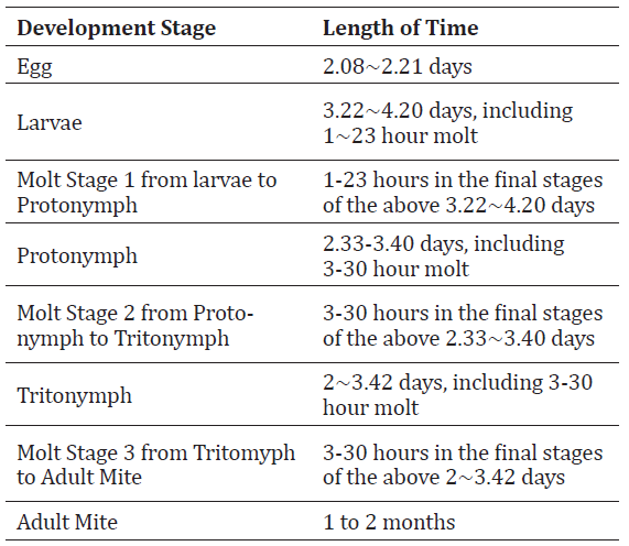
The purpose of these simulated cycles was to demonstrate the variation in the need for dose schedule length based on known and theoretical yet plausible outcomes with in vivo infections.
This shows that the timing of ivermectin treatment and molt periods are a significant factor in the number of doses required due to molt period ivermectin resistance and the mites’ potential one-third survival rates previously observed (Feng et al. 2023).
Effective doses and schedule
In all cases, in each patient, it was found that stabilising the patient and treatment of the co-morbidities was a higher priority over the Sarcoptes scabiei infection. Once treatment was started for the Sarcoptes scabiei infestation, the minimum doses were found to be between 5 and 9 doses (given as one dose per 7- day interval). The Sarcoptes scabiei resistance to ivermectin during any of the three life cycle molting processes is a likely factor. Ivermectin as a milder medication over a longer schedule was preferred over a single dose of other medication with higher toxicity risk, which did not pose a problem since the co-morbidities all required a long schedule to treat and recover from and all of the patients tolerated the captivity well since a comfortable environment was provided and patients were supervised by a care provider with experience in red fox behaviour, body language and medical care.
It was not clear during this study whether the non-responsiveness of some patients to some doses was due to resistance or due to other operational factors. It was not possible to determine with certainty whether the life cycle of the mites and the molting period resistance was a factor or whether biofilms in general came into play. All environmental factors were the same across all patients, although co-morbidities differed across many of the patients (other than juvenile siblings rescued together), and it is unclear whether there were any patient-specific biochemical factors in relation to their other indications having an effect on the ivermectin efficacy. I can only speculate the possibility of the mite molting periods playing a role, given that patients with the same or similar indications had different reactions, including same-age siblings (juveniles rescued together at similar stages of infection, for example).
While there have been a variety of treatment methods published for wildlife, there still appears to be a lack of consensus, particularly with red foxes (Rowe et al. 2019).
Other Findings:
While not part of this report, all adult red foxes in this study were found to have lungworm eggs (Eucoleus aerophilus and/or Eucoleus boehmi) in each of their faecal floats under microscopy, all of whom were asymptomatic and separately treated accordingly prior to release back to the wild.
Clinical significance:
Red foxes usually mask injury and health conditions incredibly well until extremely advanced and often signs of slow decline can be almost undetectable until final stages, sometimes within days, hours or minutes of death. This means a fox may seem fine, even with extreme injuries or conditions, until hours or minutes before passing away due to their natural survival instincts. Experience with and knowledge of the species behaviour and body language, or consulting with those who have, is essential to effectively treating and diagnosing. This can also be a reason foxes are not rescued until advanced stages of indications are obvious.
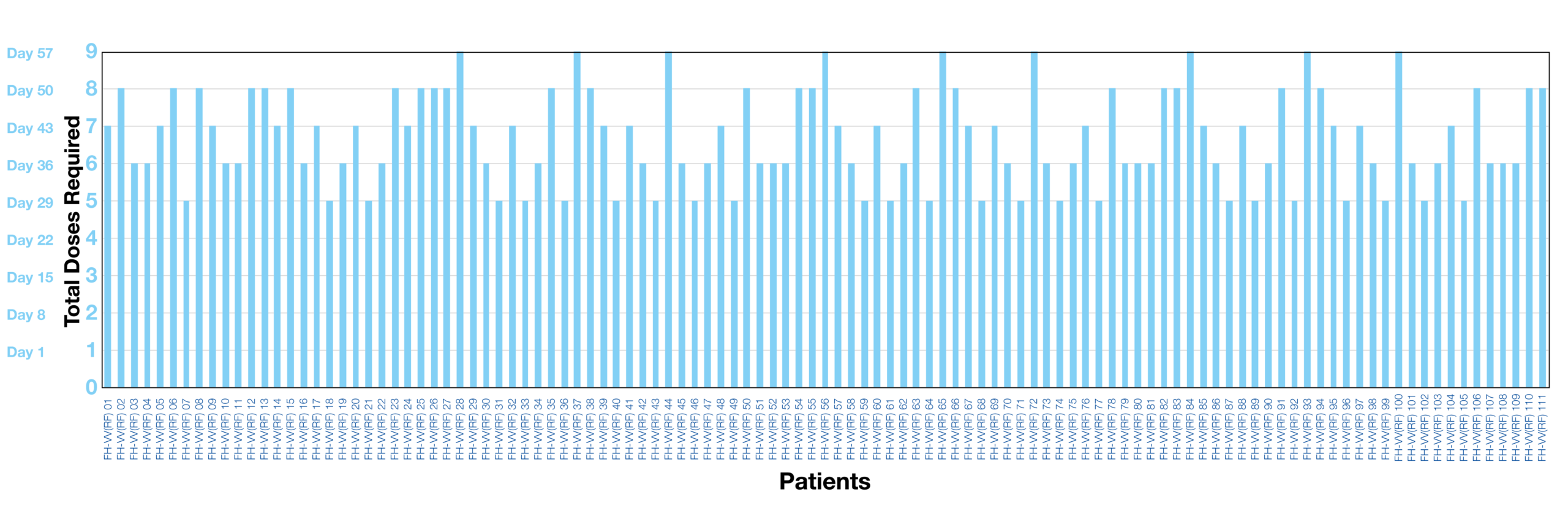
Figure 2. Summary of the number of doses of ivermectin 1% injectable w/v solution required for each individual patient to eradicate the infestation of Sarcoptes scabiei, ranging between 5 and 9 doses (given as one dose per 7-day interval).
Discussion
While ivermectin remains a common treatment, studies on newer drugs such as isoxazolines may have superior efficacy and a single-dose regimen due to longer half- life and coverage for the entire mite life cycle, and to avoid repeated handling. As this was a retrospecific case series, where patient records were selected retrospectively, the qualifying criteria for patients discussed in this paper were those being administered ivermectin specifically; therefore, a comparative study was not carried out, and those administered isoxazolines or other remedies instead were not included. It should be noted, however, that the condition red foxes come into care in these cases is usually at an advanced stage. By the time foxes are able to be seen or captured for treatment, it is usually because they have become weak, desperate, and finally showing signs of distress and disease. Red foxes are remarkably resilient acting on natural instincts for survival. They are well known among those who hold expertise in their behaviour, lifestyle and medical treatments to mask pain and medical conditions with almost absolute mastery. By the time a red fox shows symptoms, they are usually unable to mask these any longer, which means patients like these usually need to be considered urgent cases. Red foxes are not like companion animals, who may show symptoms very early on. In the cases discussed above, this means the patients were already advanced in their inability to filter out toxic loads efficiently, and so their elimination pathways were a priority area of concern and of higher priority than the Sarcoptes scabiei infection. Many of the foxes in these presented cases had a very guarded prognosis upon intake, so lessening toxic shock potential or further toxin intake was a consideration. For this reason, isoxazolines were not administered as the patients required long-term care for their comorbidities, as is often the case with Sarcoptes scabiei red fox patients. Even if isoxazolines are chosen, many of the comorbidities they have take many weeks to rectify effectively regardless of avoiding relapse after release back to the wild.
Given this, it is suggested that caution may be warranted if choosing isoxazolines due to the potential additional toxic load and the initial temporarily retarded elimination pathways, considering that comorbidities noted in many of the patients included renal, hepatic and lymphatic drainage indications with anaemia. If choosing this classification of treatment, delaying their use would be suggested, which in some cases may negate one of their main benefits of single use, particularly as some rescue centres may appear to choose them for the purpose of reducing captivity time. To the author’s knowledge, from own experience and discussions with other rehabilitators over the years, isoxazolines or other pharmaceutical or synthetic drugs may potentially cause suspected toxin-related adverse reactions or potentially fatal results if given too early, which the author has witnessed in early cases leading to a change in regimen and schedule.
In many cases, the author finds success in choosing to carry out cleaning of the patient’s skin upon intake by bathing while following handling caution such as the use of effective muzzle, proper handling without the need of bite gloves (full arm-length gloves with nitrile over gloves and disposable apron are usually sufficient and provide effective dexterity and safe handling), and the use of a veterinary tub table with a quiet warm free flowing shower attachment nozzle. A mild flea, tick and anti-fungal shampoo was used in these specific cases to address any potential topical bacterial and fungal presence. This seems to be sufficient. In some cases, a second bathing is followed up one or two weeks later on a case-by-case basis and depending on the patient’s behaviour and personality as they recover and as the skin improves with sloughing of crust and dead skin. Red foxes have a very dense double coat, so this bathing helps provide a less favourable environment for any stage of the mite life cycle.
Many foxes in cases like these where they are significantly immunocompromised are accepting of the treatment through weakness and almost limp or unreactive (as opposed to patients with minor ailment who are still able to be naturally full defensive), making this process in the first few hours of rescue fairly straight forward. No lasting mental or physical adverse effects were shown in any of the patients bathed. Some may choose to administer natural or pharmaceutical methods surrounding bacterial infection of the skin, the author has found oral pharmaceutical antibiotics to be mostly unnecessary unless there are clear indications and confirmation of specific bacterial overgrowth on wounds (such as those who came into care with bite wounds), and is assessed on a case-by-case basis. In this paper, the patients without wounds relating to other injuries were not administered pharmaceutical antibiotics, in favour of preserving the gut microbiome to aid healing. Due to the bathing stage, the author saw nothing to indicate that antibiotics were required throughout the entire treatment schedule for those admitted without infected wounds unrelated to the Sarcoptes scabiei infection (such as animal bites).
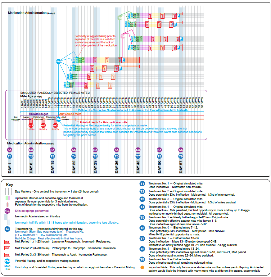
Figure 3. Simulated model of a young female mite that mated once and laid 3 eggs/day for 4 days before dying from ivermectin. Resulting offspring survived up to 3 treatments, mating and laying eggs before death; their offspring similarly did not survive beyond 3 further treatments. Despite originating from a single, short-lived female, the host still required 9 ivermectin doses at 7-day intervals to eradicate the infestation, owing to molt periods coinciding with administration days. (To download high resolution, click here)
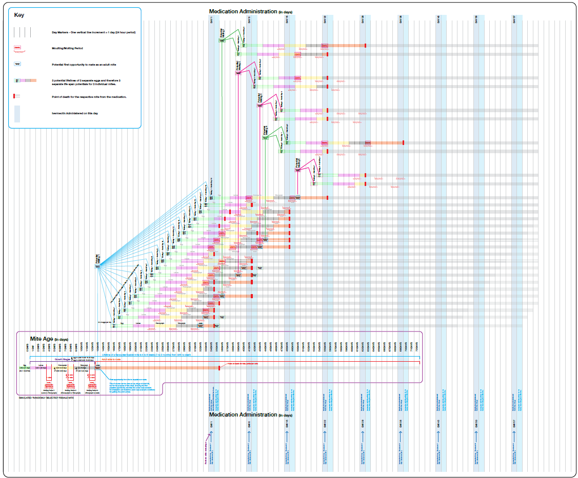
Figure 4. Simulated model of an older female mite (cf. Fig 2.) that laid 3 eggs/day for 19 consecutive days, with resulting offspring reproducing multiple times. Despite greater reproductive output than Fig 2., the host required only 6 ivermectin doses at 7-day intervals to eradicate the theoretical bloodline, owing to favourable timing of administration relative to molt periods in the original mite and her offspring. (To download high resolution, click here)
Recommendations on red fox rescue
It is the author’s opinion through treating a large number of patients like this over the years (Fox, R., unpub. data) that the treatment of Sarcoptes scabiei in the wild population of red foxes, through rescue organisations should be treated as a secondary issue and the primary issues, such as malnutrition and/or wounds rectified as a priority with the mange treated once the patient is stable and improved somewhat and their elimination pathways are functioning optimally, with full confirmation by skin scrapings (not assumptions) to confirm complete eradication of the mange, and healing of other co-morbidities (which could be between 6–10 weeks or longer before releasing back to the wild to increase chances of survival and reduce possibility of reinfection upon release. It would also be prudent to factor in the original reason for rescue and indications of the patient which regards to whether it is best practice to releasing the fox back to the exact location or to soft release carefully and effectively to another area abundant with natural resources and prey without disrupting the balance in that area, on the basis of a patient coming in with severe malnutrition and whether it would be wise to release back to an area clearly lacking in natural resources or where they may have been pushed away from available food (particularly in heavily populated urban environments with competitive resource guarding by other wildlife or red foxes) or in an area that has a known problem for mange suffering foxes. The author believes it is the responsibility of any organisation rescuing and/or rehabilitating red foxes with a view to release back to the wild upon healthy recovery to make sufficient efforts to survey the area and ensure survival chances rather than a quick turnaround of patients and releasing them to the same location for convenience. It should be noted that in the author’s experience of personal hands-on care with many foxes of different indications, red foxes generally tolerate both short and long-term veterinary captivity during treatment so long as the right enclosure, regimen and calm, supervised environment are provided, and the carers respect and understand fox-specific behaviour and body language.
TO DOWNLOAD SUPPLEMENTARY MATERIAL, CLICK HERE.
Acknowledgement
The author thanks Dr Kwint HNE, Dr Giurgiu RS, Dr Klocek M, Dr Hamilton CV, and RVN’s Williams C, Horsnell MA, Newman GN, Klein NE, and Berwick L for assistance in triage/examination of a small number of the patients in this manuscript, and Dr Lategan J and Hill DJ for kind support and assistance.
CONFLICT OF INTEREST
Ryan Fox is the founder of The Fox Hospital, where the patients were treated; however, the treatment was carried out as part of standard routine care at the charity and not for commercial purposes, so there was no financial gain, with the manuscript being written retrospectively from previous records simply reporting patient findings.
DATA AVAILABILITY
All data underlying the results, including tables and figures, are available as part of the article, and no additional source data are required.
AUTHOR CONTRIBUTION
Conceptualisation: RF.
Sample gathering, Data Curation and Formal Analysis: RF.
Investigation, Methodology and Project administration: RF.
Writing — original draft and preparation: RF.
Writing — review: RF.
Edited By
Pradeep Kumar Malik
Wildlife Institute of India, Dehradun, India.
*CORRESPONDENCE
Ryan Fox
✉ researchpapers@thefoxhospital.org
CITATION
Fox, R. (2026). Efficacy and schedule of Ivermectin in vivo against naturally acquired infestation of Sarcoptes scabiei on wild red foxes (Vulpes vulpes) treated in a controlled veterinary / clinical rehabilitation environment. Journal of Wildlife Science, 3(2), 53-61. https://doi.org/10.63033/JWLS.WIOO2051
COPYRIGHT
© 2026 Fox. This is an open-access article, immediately and freely available to read, download, and share. The information contained in this article is distributed under the terms of the Creative Commons Attribution License (CC BY 4.0
 ), allowing for unrestricted use, distribution, and reproduction in any medium, provided the original work is properly cited in accordance with accepted academic practice. Copyright is retained by the author(s).
PUBLISHED BY
Wildlife Institute of India, Dehradun, 248 001 INDIA
PUBLISHER'S NOTE
The Publisher, Journal of Wildlife Science or Editors cannot be held responsible for any errors or consequences arising from the use of the information contained in this article. All claims expressed in this article are solely those of the author(s) and do not necessarily represent those of their affiliated organisations or those of the publisher, the editors and the reviewers. Any product that may be evaluated or used in this article or claim made by its manufacturer is not guaranteed or endorsed by the publisher.
Arlian, L. G. & Vyszenski-Moher, D. L. (1988). Life cycle of Sarcoptes scabiei var. canis. The Journal of Parasitology, 74(3), 427–430. https://doi.org/10.2307/3282050
Arlian, L. G., Runyan, R. A., Achar, S. & Estes, S. A. (1984). Survival and infectivity of Sarcoptes scabiei var. canis and var. hominis. Journal of the American Academy of Dermatology, 11(2 Part 1), 210–215. https://doi.org/10.1016/S0190-9622(84)70151-4
Allerton, F., Batchelor, D., Bexfield, N., Chan, D. L., Featherstone, H., Frowde, P., Helm, J., Hodgkiss-Geere, H., Kent, A. et al. (2020). BSAVA Small Animal Formulary (10th ed., Part A: Canine and Feline). BSAVA. pp.6–7, 164–165, 172–173, 216, 369.
Bernigaud, C., Fang, F., Fischer, K., Lespine, A., Aho, L. S., Dreau, D., Kelly, A., Sutra, J. F., Moreau, F. et al. (2016). Preclinical Study of Single-Dose Moxidectin, a New Oral Treatment for Scabies: Efficacy, Safety, and Pharmacokinetics Compared to Two-Dose Ivermectin in a Porcine Model. PLoS Neglected Tropical Diseases, 10(10), e0005030. https://doi.org/10.1371/journal.pntd.0005030
Bernigaud, C., Fang, F., Fischer, K., Lespine, A., Aho, L. S., Mullins, A. J., Tecle, B., Kelly, A., Sutra, J. F. et al. (2018). Efficacy and Pharmacokinetics Evaluation of a Single Oral Dose of Afoxolaner against Sarcoptes scabiei in the Porcine Scabies Model for Human Infestation. Antimicrobial Agents and Chemotherapy, 62(9), e02334-17. https://doi.org/10.1128/aac.02334-17
CDC (2023). Clinical Care of Scabies. U.S. Centers for Disease Control and Prevention. https://www.cdc.gov/scabies/hcp/clinical-care/index.html [February 2025]
Chitty, J., Hedley, J., Saunders, R., Hildgoose, W. H. & Wissink-Argilaga, N. (2020). BSAVA Small Animal Formulary (10th ed., Part B: Exotic Pets). BSAVA. pp.169–170.
Crump, A. & Ōmura, S. (2011). Ivermectin, "wonder drug" from Japan: the human use perspective. Proceedings of the Japan Academy, Series B, 87(2), 13–28. https://doi.org/10.2183/pjab.87.13
Currie, B. J., Harumal, P., McKinnon, M. & Walton, S. F. (2004). First documentation of in vivo and in vitro ivermectin resistance in Sarcoptes scabiei. Clinical Infectious Diseases, 39(1), e8–e12. https://doi.org/10.1086/421776
Feng, S., Shi, M., Yin, Z., Di, W., Guillot, J. & Fang, F. (2023). Can Ivermectin kill Sarcoptes scabiei during the molting process? PLoS Neglected Tropical Diseases, 17(5), e0011337. https://doi.org/10.1371/journal.pntd.0011337
Furnival-Adams, J., Kiuru, C., Sagna, A. B., Mouline, K., Maia, M. & Chaccour, C. (2024). Ivermectin resistance mechanisms in ectoparasites: a scoping review. Parasitology Research, 123(5), 221. https://doi.org/10.1007/s00436-024-08223-z
Kaplan, R. M. (2004). Drug resistance in nematodes of veterinary importance: a status report. Trends in Parasitology, 20(10), 477–481. https://doi.org/10.1016/j.pt.2004.08.001
Laing, R., Gillan, V. & Devaney, E. (2017). Ivermectin—Old Drug, New Tricks? Trends in Parasitology, 33(6), 463–472. https://doi.org/10.1016/j.pt.2017.02.004
Mounsey, K. E., Walton, S. F., Innes, A., Cash-Deans, S. & McCarthy, J. S. (2017). In Vitro Efficacy of Moxidectin versus Ivermectin against Sarcoptes scabiei. Antimicrobial Agents and Chemotherapy, 61(8), e00381-17. https://doi.org/10.1128/AAC.00381-17
NHS (2024). Clinical Guideline: Scabies Therapeutic Management, Acute & Primary Care. National Health Service. https://rightdecisions.scot.nhs.uk/media/q3cnc1dq/1179-scabies-therapeutic-management-acute-primary-care-v1.pdf [February 2025]
Rowe, M. L., Whiteley, P. L. & Carver, S. (2019). The treatment of sarcoptic mange in wildlife: a systematic review. Parasites & Vectors, 12(1), 99. https://doi.org/10.1186/s13071-019-3340-z
Scott, D. M., Baker, R., Tomlinson, A., Berg, M. J., Charman, N. & Tolhurst, B. A. (2020). Spatial distribution of sarcoptic mange (Sarcoptes scabiei) in urban foxes (Vulpes vulpes) in Great Britain as determined by citizen science. Urban Ecosystems 23, 1127–1140. https://doi.org/10.1007/s11252-020-00985-5
Shoop, W. L. (1993). Ivermectin resistance. Parasitology Today, 9(5), 154–159. https://doi.org/10.1016/0169-4758(93)90136-4
Soulsbury, C. D., Iossa, G., Baker, P. J., Cole, N. C., Funk, S.M. & Harris, S. (2007). The impact of sarcoptic mange Sarcoptes scabiei on the British fox Vulpes vulpes population. Mammal Review, 37, 278-296. https://doi.org/10.1111/j.1365-2907.2007.00100.x

Edited By
Pradeep Kumar Malik
Wildlife Institute of India, Dehradun, India.
*CORRESPONDENCE
Ryan Fox
✉ researchpapers@thefoxhospital.org
CITATION
Fox, R. (2026). Efficacy and schedule of Ivermectin in vivo against naturally acquired infestation of Sarcoptes scabiei on wild red foxes (Vulpes vulpes) treated in a controlled veterinary / clinical rehabilitation environment. Journal of Wildlife Science, 3(2), 53-61. https://doi.org/10.63033/JWLS.WIOO2051
COPYRIGHT
© 2026 Fox. This is an open-access article, immediately and freely available to read, download, and share. The information contained in this article is distributed under the terms of the Creative Commons Attribution License (CC BY 4.0), allowing for unrestricted use, distribution, and reproduction in any medium, provided the original work is properly cited in accordance with accepted academic practice. Copyright is retained by the author(s).
PUBLISHED BY
Wildlife Institute of India, Dehradun, 248 001 INDIA
PUBLISHER'S NOTE
The Publisher, Journal of Wildlife Science or Editors cannot be held responsible for any errors or consequences arising from the use of the information contained in this article. All claims expressed in this article are solely those of the author(s) and do not necessarily represent those of their affiliated organisations or those of the publisher, the editors and the reviewers. Any product that may be evaluated or used in this article or claim made by its manufacturer is not guaranteed or endorsed by the publisher.
Arlian, L. G. & Vyszenski-Moher, D. L. (1988). Life cycle of Sarcoptes scabiei var. canis. The Journal of Parasitology, 74(3), 427–430. https://doi.org/10.2307/3282050
Arlian, L. G., Runyan, R. A., Achar, S. & Estes, S. A. (1984). Survival and infectivity of Sarcoptes scabiei var. canis and var. hominis. Journal of the American Academy of Dermatology, 11(2 Part 1), 210–215. https://doi.org/10.1016/S0190-9622(84)70151-4
Allerton, F., Batchelor, D., Bexfield, N., Chan, D. L., Featherstone, H., Frowde, P., Helm, J., Hodgkiss-Geere, H., Kent, A. et al. (2020). BSAVA Small Animal Formulary (10th ed., Part A: Canine and Feline). BSAVA. pp.6–7, 164–165, 172–173, 216, 369.
Bernigaud, C., Fang, F., Fischer, K., Lespine, A., Aho, L. S., Dreau, D., Kelly, A., Sutra, J. F., Moreau, F. et al. (2016). Preclinical Study of Single-Dose Moxidectin, a New Oral Treatment for Scabies: Efficacy, Safety, and Pharmacokinetics Compared to Two-Dose Ivermectin in a Porcine Model. PLoS Neglected Tropical Diseases, 10(10), e0005030. https://doi.org/10.1371/journal.pntd.0005030
Bernigaud, C., Fang, F., Fischer, K., Lespine, A., Aho, L. S., Mullins, A. J., Tecle, B., Kelly, A., Sutra, J. F. et al. (2018). Efficacy and Pharmacokinetics Evaluation of a Single Oral Dose of Afoxolaner against Sarcoptes scabiei in the Porcine Scabies Model for Human Infestation. Antimicrobial Agents and Chemotherapy, 62(9), e02334-17. https://doi.org/10.1128/aac.02334-17
CDC (2023). Clinical Care of Scabies. U.S. Centers for Disease Control and Prevention. https://www.cdc.gov/scabies/hcp/clinical-care/index.html [February 2025]
Chitty, J., Hedley, J., Saunders, R., Hildgoose, W. H. & Wissink-Argilaga, N. (2020). BSAVA Small Animal Formulary (10th ed., Part B: Exotic Pets). BSAVA. pp.169–170.
Crump, A. & Ōmura, S. (2011). Ivermectin, "wonder drug" from Japan: the human use perspective. Proceedings of the Japan Academy, Series B, 87(2), 13–28. https://doi.org/10.2183/pjab.87.13
Currie, B. J., Harumal, P., McKinnon, M. & Walton, S. F. (2004). First documentation of in vivo and in vitro ivermectin resistance in Sarcoptes scabiei. Clinical Infectious Diseases, 39(1), e8–e12. https://doi.org/10.1086/421776
Feng, S., Shi, M., Yin, Z., Di, W., Guillot, J. & Fang, F. (2023). Can Ivermectin kill Sarcoptes scabiei during the molting process? PLoS Neglected Tropical Diseases, 17(5), e0011337. https://doi.org/10.1371/journal.pntd.0011337
Furnival-Adams, J., Kiuru, C., Sagna, A. B., Mouline, K., Maia, M. & Chaccour, C. (2024). Ivermectin resistance mechanisms in ectoparasites: a scoping review. Parasitology Research, 123(5), 221. https://doi.org/10.1007/s00436-024-08223-z
Kaplan, R. M. (2004). Drug resistance in nematodes of veterinary importance: a status report. Trends in Parasitology, 20(10), 477–481. https://doi.org/10.1016/j.pt.2004.08.001
Laing, R., Gillan, V. & Devaney, E. (2017). Ivermectin—Old Drug, New Tricks? Trends in Parasitology, 33(6), 463–472. https://doi.org/10.1016/j.pt.2017.02.004
Mounsey, K. E., Walton, S. F., Innes, A., Cash-Deans, S. & McCarthy, J. S. (2017). In Vitro Efficacy of Moxidectin versus Ivermectin against Sarcoptes scabiei. Antimicrobial Agents and Chemotherapy, 61(8), e00381-17. https://doi.org/10.1128/AAC.00381-17
NHS (2024). Clinical Guideline: Scabies Therapeutic Management, Acute & Primary Care. National Health Service. https://rightdecisions.scot.nhs.uk/media/q3cnc1dq/1179-scabies-therapeutic-management-acute-primary-care-v1.pdf [February 2025]
Rowe, M. L., Whiteley, P. L. & Carver, S. (2019). The treatment of sarcoptic mange in wildlife: a systematic review. Parasites & Vectors, 12(1), 99. https://doi.org/10.1186/s13071-019-3340-z
Scott, D. M., Baker, R., Tomlinson, A., Berg, M. J., Charman, N. & Tolhurst, B. A. (2020). Spatial distribution of sarcoptic mange (Sarcoptes scabiei) in urban foxes (Vulpes vulpes) in Great Britain as determined by citizen science. Urban Ecosystems 23, 1127–1140. https://doi.org/10.1007/s11252-020-00985-5
Shoop, W. L. (1993). Ivermectin resistance. Parasitology Today, 9(5), 154–159. https://doi.org/10.1016/0169-4758(93)90136-4
Soulsbury, C. D., Iossa, G., Baker, P. J., Cole, N. C., Funk, S.M. & Harris, S. (2007). The impact of sarcoptic mange Sarcoptes scabiei on the British fox Vulpes vulpes population. Mammal Review, 37, 278-296. https://doi.org/10.1111/j.1365-2907.2007.00100.x


